Home
/ Shoulder Tendon And Ligament Anatomy - Tendons vs. ligaments: What they are, injuries, and treatments : The distal joint between the tibia and fibula is an example of a.
Shoulder Tendon And Ligament Anatomy - Tendons vs. ligaments: What they are, injuries, and treatments : The distal joint between the tibia and fibula is an example of a.
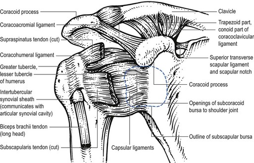
Shoulder Tendon And Ligament Anatomy - Tendons vs. ligaments: What they are, injuries, and treatments : The distal joint between the tibia and fibula is an example of a.. Anteriorly the subscapularis tendon is separated from the supraspinatus tendon by a gap, the rotator interval, which these ligaments pass from the coracoid and glenoid respectively, and insert into the humeral head on either. Corey chakarun from shin imaging in california. The flexor digitorum superficialis (fds) and flexor digitorum. The patellar tendon on the front of the knee is part of the quadriceps mechanism. Learn about the muscles, tendons, bones, and ligaments that comprise the knee joint anatomy.
More about dental anatomy and periodontal ligaments you can find in the article about the anatomy of the teeth and this interesting video tutorial. Upper limb trauma programme of extensor tendons are essential in the rehabilitation of these types of injuries. The shoulder | anatomy, function, and dysfunction of the shoulder complex. Instead of your doctor simply saying that the patient knee hurts, he or. Tendons, ligaments, bone, and cartilage are connective tissues in which the activities of various cellular populations are responsible for synthesis and maintenance of large amounts of extracellular matrix that should, theoretically, be dynamically optimized to respond to mechanical demands.
Shoulder Tendon And Ligament Anatomy - PPT - The ... from musculoskeletalkey.com Shoulder joint is formed by a group of ligaments that connect humerus to glenoid. The coracohumeral ligament strengthens the capsule from above and stretches from the root of the coracoid process to the greater tuberosity • the tendons of these muscles are fused to the underlying capsule of the shoulder. Tendons and ligaments are complex structures and have different anatomical and dynamic properties. A joint capsule is a watertight sac that surrounds a joint. (3) a syndesmosis is a joint in which a ligament connects two bones, allowing for a little movement (amphiarthroses). Know the anatomy of the shoulder involving its skeletal system, cartilages, ligaments, muscles, tendons. Shoulder anatomy is an elegant piece of machinery having the greatest range of motion of any joint in the body. The human shoulder is made up of three bones:
The shoulder joint (glenohumeral joint) is a ball and socket joint between the scapula and the humerus.
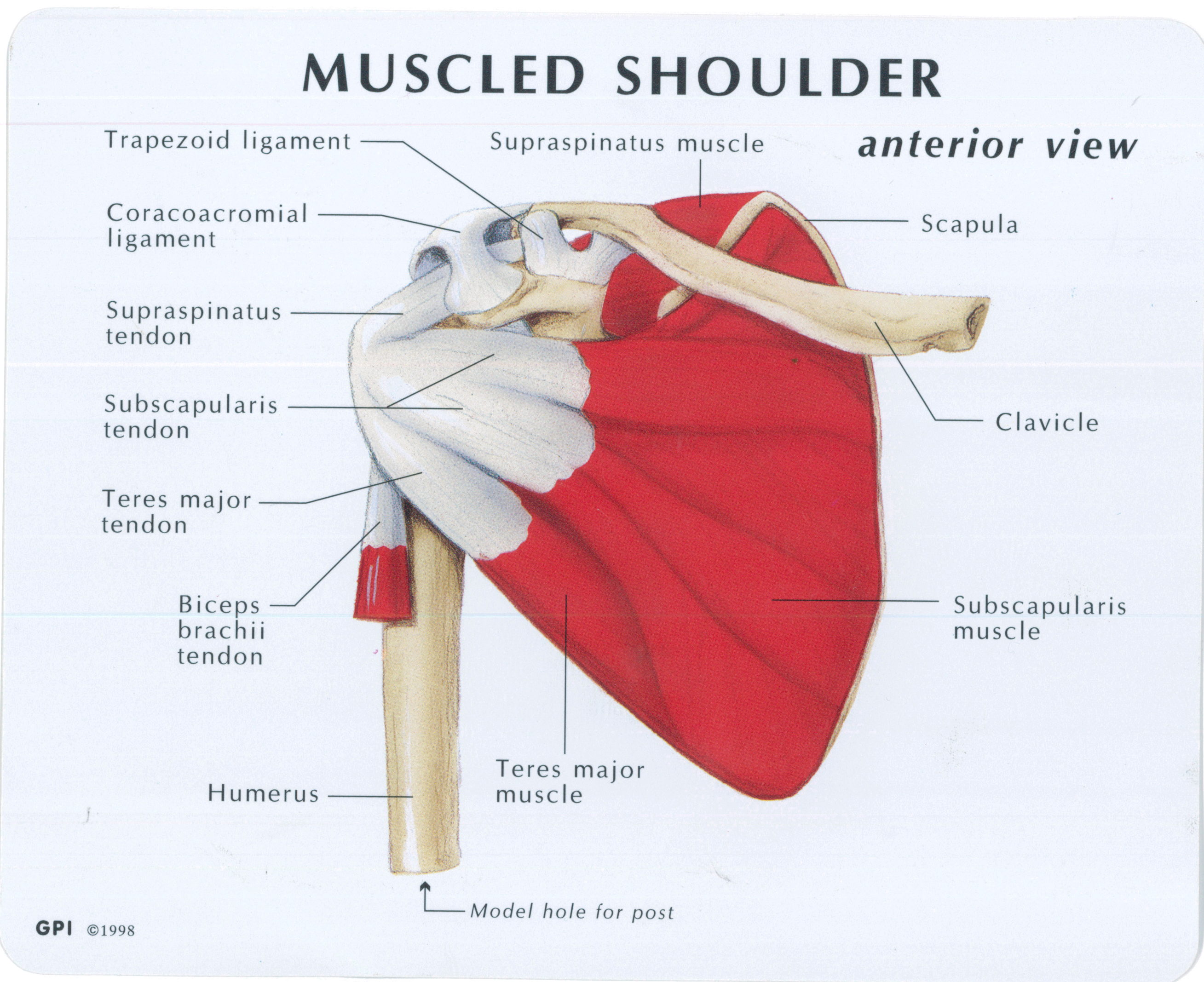
Although scarring depends on the quality and quantity of the injured tissues, it can be. Other smaller muscles and tendons surround the knee joint as well. More about dental anatomy and periodontal ligaments you can find in the article about the anatomy of the teeth and this interesting video tutorial. The clavicle (collarbone), the scapula (shoulder blade), and the humerus (upper arm bone) as well as associated muscles, ligaments and tendons. The four tendons of these muscles converge to form the rotator cuff tendon. Start studying shoulder ligaments and tendons. Muscles allow us to move. Ligaments are soft tissue structures that connect bones to bones. The ca ligament along with the acromial process create the outlet of the shoulder thru which passes the supraspinatus tendon of the rotator cuff. Ball and socket joint between the head of the humerus and the glenoid joint: Thickening or calcium deposits in the supraspinatus tendon or subacromial bursitis results in pain during abduction of shoulder joint from 60° to 120°. (3) a syndesmosis is a joint in which a ligament connects two bones, allowing for a little movement (amphiarthroses). Shoulder joint allows lifting, pushing and pulling by upper extremity.
Unlike tendon and ligament, cartilage can withstand a great degree of compression. Joints are bone such as knee, hip, and shoulder that connect in different ways to allow you to kneel, run or lift your arm up. Ligaments are soft tissue structures that connect bones to bones. These ligaments are main source of stability for the shoulder. Tendons, ligaments, bone, and cartilage are connective tissues in which the activities of various cellular populations are responsible for synthesis and maintenance of large amounts of extracellular matrix that should, theoretically, be dynamically optimized to respond to mechanical demands.
Muscled Shoulder Joint Model - MedWest Medical Supplies from www.medwest.ca (3) a syndesmosis is a joint in which a ligament connects two bones, allowing for a little movement (amphiarthroses). The shoulder joint (glenohumeral joint) is a ball and socket joint between the scapula and the humerus. Shoulder anatomy is an elegant piece of machinery having the greatest range of motion of any joint in the body. In order to achieve this flexibility but maintain a stable shoulder, there is a complex interplay between the joints, muscles and ligaments. In plain english, it is gristle. More about dental anatomy and periodontal ligaments you can find in the article about the anatomy of the teeth and this interesting video tutorial. Ligaments are soft tissue structures that connect bones to bones. Arm flexion, extension, adduction, abduction, and internal and external rotation blood.
Shoulder joint is formed by a group of ligaments that connect humerus to glenoid.
(3) a syndesmosis is a joint in which a ligament connects two bones, allowing for a little movement (amphiarthroses). Shoulder joint allows lifting, pushing and pulling by upper extremity. Other smaller muscles and tendons surround the knee joint as well. Superior glenohumeral ligament and coracohumeral ligament are the primary restraints to posterior translation with the are flexed, adducted and prevents inferior translation and external rotation in the abducted shoulder, and provides stability to the long head of the biceps tendon (neer cs ii, corr. The four tendons of these muscles converge to form the rotator cuff tendon. Although scarring depends on the quality and quantity of the injured tissues, it can be. Joints can be grouped by their structure into fibrous, cartilaginous, and synovial joints. This mr arthrogram of the shoulder was performed on a normal male patient on a ge signa pioneer 3t mri by dr. Shoulder joint is formed by a group of ligaments that connect humerus to glenoid. Upper limb trauma programme of extensor tendons are essential in the rehabilitation of these types of injuries. The shoulder joint (glenohumeral joint) is a ball and socket joint between the scapula and the humerus. The shoulder is comprised of a ball (humerus) and socket (scapula), bones, ligaments, tendons and muscles that move the arms and connect them to the torso. The knee joint has 3 pieces, the hip joint has 2 pieces, the.
More about dental anatomy and periodontal ligaments you can find in the article about the anatomy of the teeth and this interesting video tutorial. Tendons, ligaments, bone, and cartilage are connective tissues in which the activities of various cellular populations are responsible for synthesis and maintenance of large amounts of extracellular matrix that should, theoretically, be dynamically optimized to respond to mechanical demands. Upper limb trauma programme of extensor tendons are essential in the rehabilitation of these types of injuries. In plain english, it is gristle. In order to achieve this flexibility but maintain a stable shoulder, there is a complex interplay between the joints, muscles and ligaments.
Male Shoulder Ligaments And Biceps Muscles Isolated In ... from media.istockphoto.com (1) the collagen fibers are closely packed (dense) and leave relatively little open space, and (2) the fibers are parallel to each other (regular). (3) a syndesmosis is a joint in which a ligament connects two bones, allowing for a little movement (amphiarthroses). Sometimes, the overuse and repetitive motion of our body parts such as hands, biceps, shoulder, ankles and knees cause problems like tendonitis. Learn vocabulary, terms and more with flashcards, games and other study tools. Tendons and ligaments are complex structures and have different anatomical and dynamic properties. The four tendons of these muscles converge to form the rotator cuff tendon. Joints can be grouped by their structure into fibrous, cartilaginous, and synovial joints. These tendinous insertions along with the articular capsule, the coracohumeral ligament, and the joint capsule is a lax fibrous sheath extends from the anatomical neck of the humerus to the border of the glenoid fossa.
Both tendons and ligaments are dense regular connective tissue, because of its two properties:
Unlike tendon and ligament, cartilage can withstand a great degree of compression. The system of ligaments in the vertebral column, combined with the tendons and muscles, provides a natural brace to help protect the spine from injury. These ligaments are main source of stability for the shoulder. The distal joint between the tibia and fibula is an example of a. Muscles allow us to move. Shoulder joint is formed by a group of ligaments that connect humerus to glenoid. Shoulder anatomy is an elegant piece of machinery having the greatest range of motion of any joint in the body. This is known as 'painful arc syndrome'. Sometimes, the overuse and repetitive motion of our body parts such as hands, biceps, shoulder, ankles and knees cause problems like tendonitis. The human shoulder is made up of three bones: Superior glenohumeral ligament and coracohumeral ligament are the primary restraints to posterior translation with the are flexed, adducted and prevents inferior translation and external rotation in the abducted shoulder, and provides stability to the long head of the biceps tendon (neer cs ii, corr. Tendons and ligaments are complex structures and have different anatomical and dynamic properties. Both tendons and ligaments are dense regular connective tissue, because of its two properties:
The human shoulder is made up of three bones: shoulder tendon anatomy. These tendinous insertions along with the articular capsule, the coracohumeral ligament, and the joint capsule is a lax fibrous sheath extends from the anatomical neck of the humerus to the border of the glenoid fossa.
